WEBINAR | Up to 40% Faster Automated TEM Lamella Preparation with new Ga⁺ FIB Column

Investigate fungal infection pathways in plants with Cryo-SEM on Tescan CLARA

Visualize plant vascular ultrastructure, infection spread, and host response using Cryo-SEM imaging for fungal infection analysis.

hero
hero
Unlocked content

Visualize Pathogen Spread in Ash Tree Vessels with Cryo-SEM

Understanding how plant diseases originate and progress, particularly in their early stages, is essential for developing effective control and treatment strategies. In agriculture and forestry, such insights are critical to mitigating yield losses caused by pathogenic fungi.

Fungal infections in plants begin with subtle yet critical ultrastructural changes. At the cellular level, compromised cell walls weaken vessel integrity, allowing pathogens to spread rapidly through plant tissues.

Conventional preparation techniques—such as chemical fixation and heavy-metal staining—can alter and distort delicate structures, making early interactions between host and pathogen difficult to observe, and are often time-consuming.

Tescan CLARA, equipped with an integrated cryo stage, enables cryogenic imaging of plant tissues in the frozen-hydrated state. This setup preserves fungal hyphae and plant vessels integrity, allowing researchers to trace infection pathways and study plant–pathogen interactions without introducing sample distortion.

Why Study Plant–Pathogen Interactions

with Tescan CLARA?

01
Root of the Problem

Conventional electron microscopy requires dehydration and chemical fixation, often causing artifacts, shrinkage, or collapse of delicate biological structures. These distortions make it difficult to study natural morphology, especially in hydrated plant tissues where fine surface details are essential to understanding infection processes.

Cryo-SEM imaging eliminates these limitations by preserving samples in a frozen-hydrated state, maintaining their native structure and spatial integrity. This makes it a powerful tool for examining plant–pathogen interactions in their true physiological context.

Tescan CLARA, equipped with an integrated cryo stage, provides the stability and resolution needed to image infected plant tissues without structural distortion. Researchers can observe early fungal colonization and trace infection pathways with exceptional clarity and depth of focus, benefiting from key Cryo-SEM advantages such as:

  • High-resolution Cryo-SEM for studying plant–pathogen interactions

  • Excellent contrast and depth of focus reveal complex tissue architecture

  • Stable cryo conditions preserve native morphology

  • Ideal for agricultural and botanical research

Investigate the progression of fungal infections in plant tissues with the Tescan CLARA, capturing early colonization and preserving native structural integrity.

02
Materials and Methods

How Plant–Pathogen Interactions Were Studied Using Tescan CLARA

Infected ash tree samples showing symptoms of fungal infection were collected and cryo-fixed using plunge freezing in liquid nitrogen. This rapid freezing process preserved tissue natural hydration and internal structure, preventing collapse or alteration for imaging.

Frozen samples were then freeze-fractured to expose xylem, cortex, and other internal regions, particularly the wood vessels that are inter-grown with the fungal hyphae and transferred to the Tescan CLARA.

Imaging was carried out with the Tescan CLARA UHR-SEM under cryogenic conditions using a low accelerating voltage (2 keV), which is ideal for preservation of delicate fungal hyphae and plant tissues.

Stable cryogenic conditions and high-resolution optics enabled detailed visualization of infection pathways while maintaining the native morphology and structural integrity of the sample.

03
Results and Discussion

Cryo Imaging of Fungal Infections in Intact Plant Tissues

Cryo-SEM imaging with Tescan CLARA enabled ultra-high resolution observation of infected ash tissues in their frozen-hydrated state, preserving both plant and fungal morphology. Colonization pathways within xylem vessels and cortical tissues were revealed without the collapse or deformation caused by traditional SEM sample preparation.

Fungal hyphae and areas of plant tissue degradation were captured with enhanced depth of focus and high contrast under low beam energy conditions.

Freeze-fractured surfaces exposed intact internal features, allowing researchers to track infection spread across layers of plant tissue. The observations provided a more accurate understanding of how fungal infection progresses within the host.

This workflow delivered consistent, detailed insight into plant–pathogen interactions, supporting the development of more accurate models of disease progression and treatment.

GET IN Touch

Contact us

Get the most out of Tescan

This is more than information; it's an advantage. We've compiled our technical whitepapers, detailed product flyers, and on-demand webinars to provide you with the knowledge that makes a real difference. Sign up now to access the insights you need to make an impact.

Tescan Instruments & Technology

Used in This Workflow

Tescan CLARA

Tescan CLARA delivers ultra-high resolution SEM imaging of frozen-hydrated samples to preserve biological structure and integrity during analysis.

Combining BrightBeam™ optics, advanced low-kV imaging, and a fully integrated cryogenic workflow, CLARA provides the stability and contrast needed to visualize delicate ultrastructures in their native state.

  • Ultra-high-resolution imaging – Capture fine ultrastructural details of hydrated and beam-sensitive biological samples.

  • BrightBeam™ electron optics – Provide outstanding resolution and contrast, even at ultra-low accelerating voltages and beam currents.

  • Streamlined cryo workflow – Integrated cryo stage and stable sample transfer minimize contamination and simplify operation.

  • Expanded field of view – Quickly navigate and locate regions of interest on large samples with Wide Field Optics™

  • Multi-user support – Manage multiple operators and experimental workflows efficiently with Tescan Essence SW

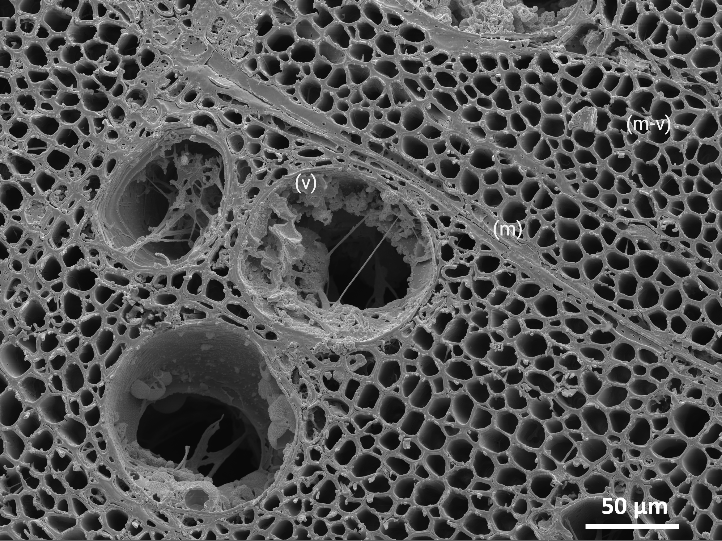
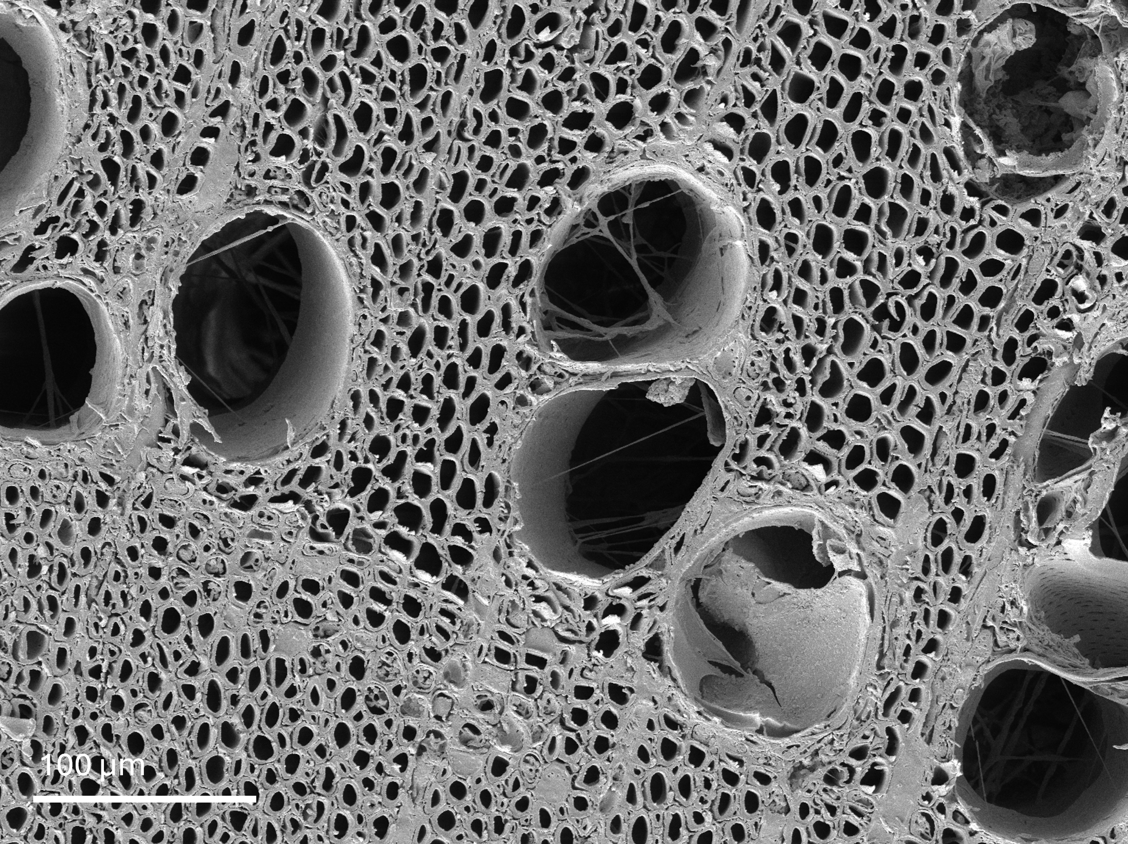
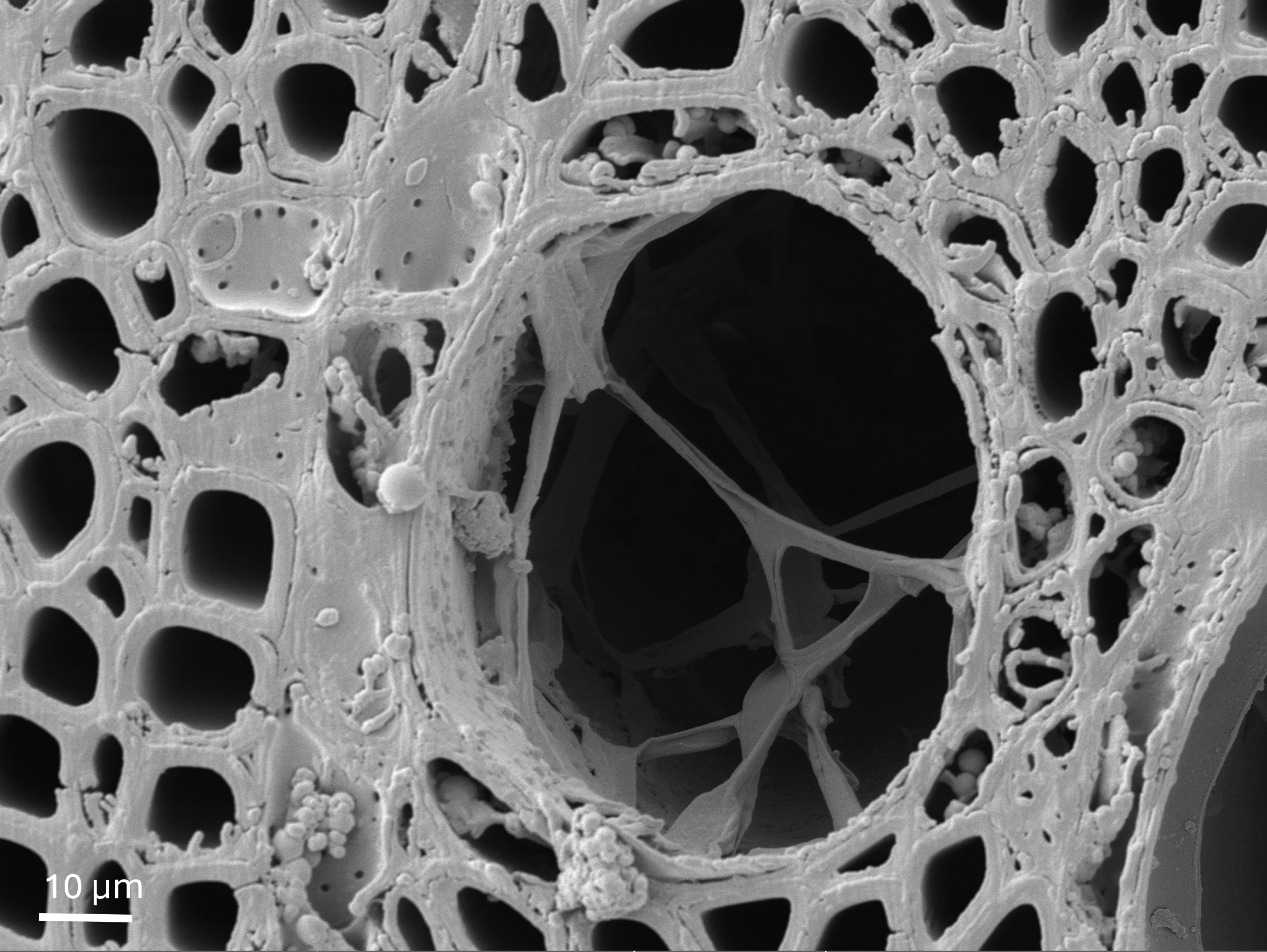
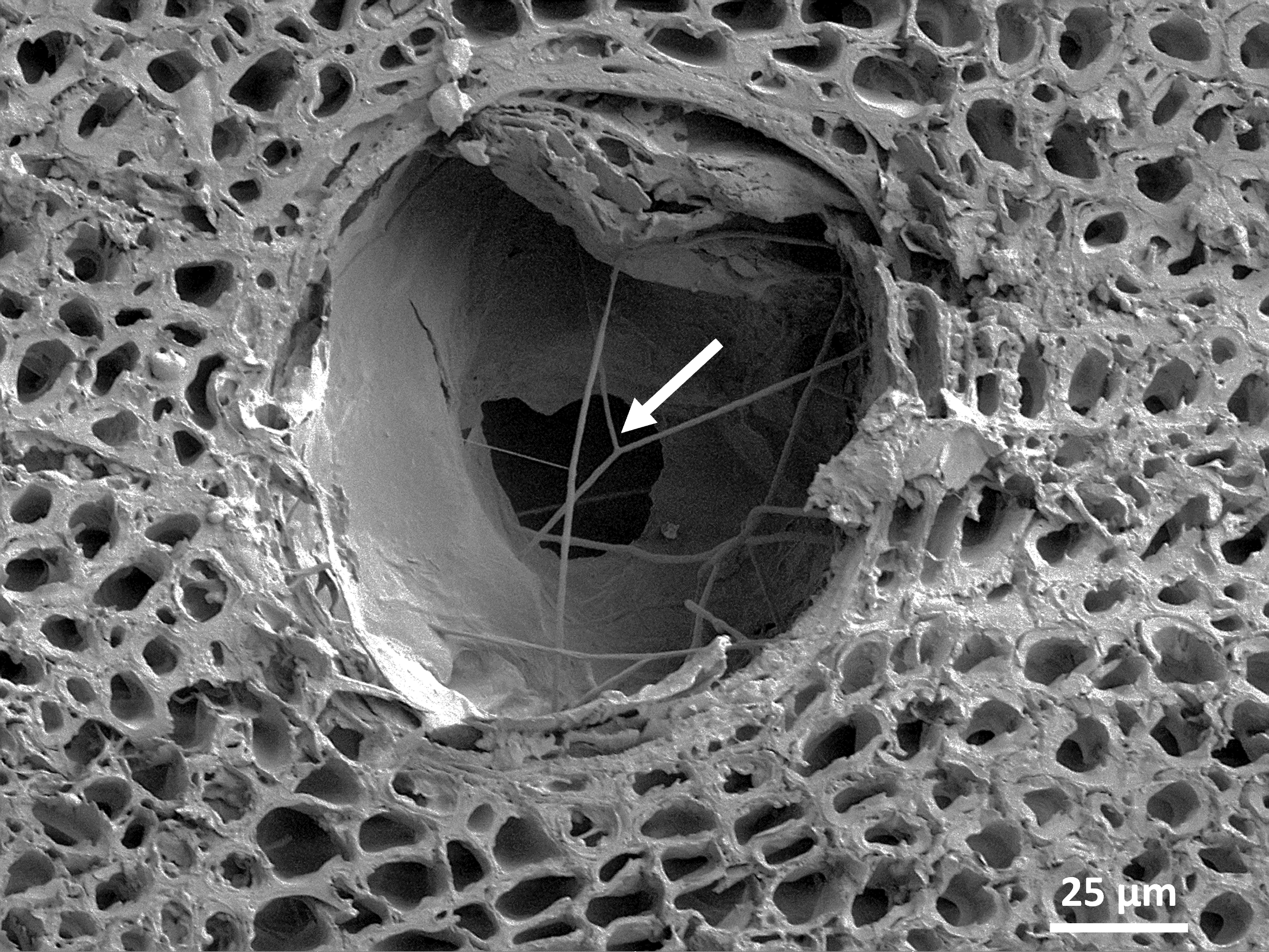
With Tescan CLARA, complex biological structures can be visualized with precision, preserving natural morphology and ensuring consistent imaging quality.

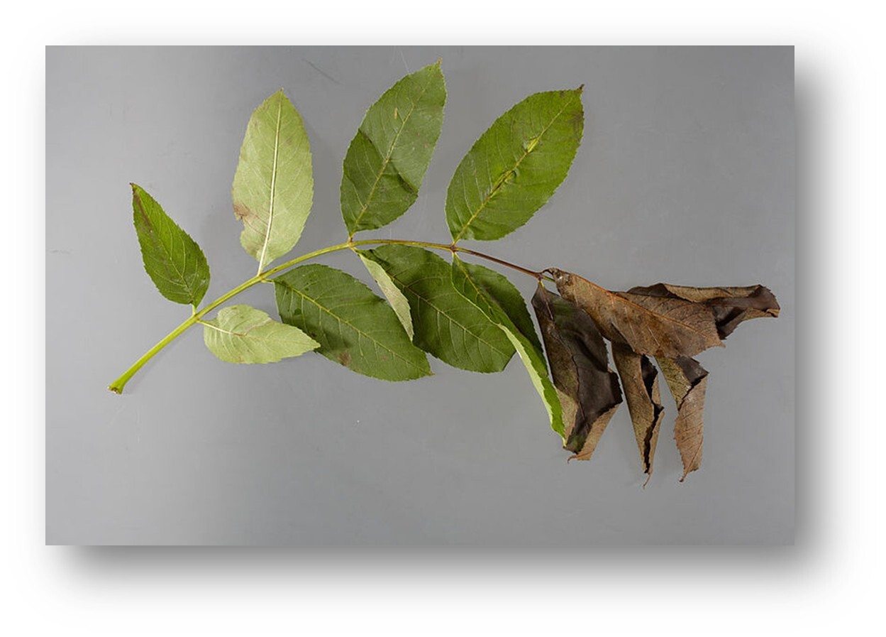
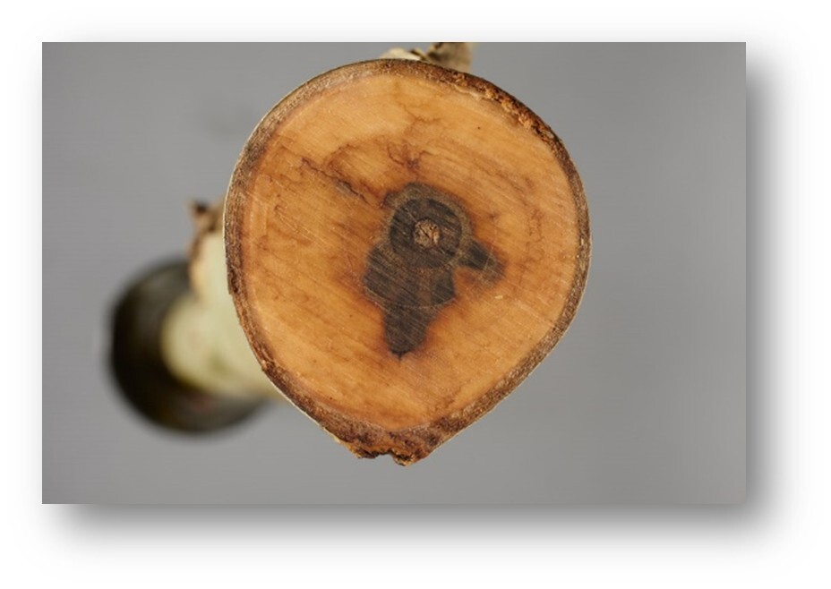
The analysis was performed in cooperation with Dr. Miroslava Mamoňová, Faculty of Wood Sciences and Technology, TU in Zvolen, Slovakia.

CLARA GM Mat. Science

GET IN Touch

Contact us

map

Where can you find us:

Tescan Brno
Libušina třída 21
623 00 Brno
Czech Republic

info@Tescan.com