WEBINAR | Up to 40% Faster Automated TEM Lamella Preparation with new Ga⁺ FIB Column

Achieve Large-Volume FIB-SEM Tomography in a Single Acquisition

Map internal features of zebrafish embryos with Xe plasma FIB-SEM tomography, capturing early development in a single, uninterrupted run without compromise.

hero
hero
Unlocked content

Reveal Embryonic Development in Zebrafish with Fast, Large-Volume Xe Plasma FIB-SEM

Zebrafish (Danio rerio)  are a key model in biological research. Their rapid development, transparent embryos, and genetic similarity to humans make them highly relevant for developmental biology, toxicology, and disease studies. Researchers can observe organogenesis in real time and later reconstruct tissue ultrastructure in 3D, gaining powerful insights into fundamental processes.

Reconstructing large biological volumes at high resolution remains challenging. Conventional Ga FIBs are slow, limited by low milling rates, and require periodic ion source reconditioning during long acquisitions due to gallium depletion. As a result, imaging often consists of fragments, leading to stitching errors and loss of context. Therefore a faster, more robust approach is needed.

With Tescan’s Xe plasma FIB-SEM, imaging continuity is no longer a barrier. Entire embryo regions can be captured in a single run, preserving spatial coherence and structural context across complex tissues.

Why Xe Plasma FIB-SEM should be your choice

for 3D Tomography?

01
Root of the Problem

Why Gallium FIB-SEM Falls Short for Large-Volume Embryo Imaging

High-resolution imaging of zebrafish embryos requires more than fine detail — it demands continuity across large volumes. Traditional gallium-based FIB-SEM systems often force imaging in fragments, leading to stitching errors and loss of context.

When studying complex tissues such as vasculature and mesenchyme, any break in acquisition can compromise data integrity. Consistent, artifact-free imaging over long runs is essential.

Tescan’s Xe plasma FIB-SEM was designed to meet these challenges, offering:

  • A stable Xe plasma ion source for continuous long-run acquisitions without downtime

  • High milling rates to reconstruct large specimen volumes efficiently

  • Consistent image quality across extended acquisitions

Combined with immersion optics for ultra-high resolution, this workflow enables imaging of entire embryo regions in a single continuous scan while preserving the structural integrity needed for accurate 3D reconstruction.

02
Materials and Methods

How Xe Plasma FIB-SEM Enables Continuous 3D Embryo Reconstruction

Zebrafish embryos (Danio rerio ) were chemically fixed, stained and embedded in resin for further analysis in Xe plasma FIB-SEM.

Regions of interest were located in the trunk, extending from the urogenital opening toward the head and encompassing the dorsal aorta and caudal vein.

FIB-SEM tomograohy was performed using a Tescan SOLARIS X2, Xe Plasma FIB-SEM system with immersion optics. A protective mask layer was applied over the surface. Volume acquisition was carried out in a single acquisition, enabling precise reconstruction of both regions with consistent surface quality and nanometer resolution.

03
Results and Discussion

Nanometer-Scale Detail and Anatomical Continuity in Zebrafish Embryo FIB-SEM Tomography

The regions of interest, the dorsal aorta with its surrounding vasculature and the mesenchymal floor, were acquired in a single, uninterrupted imaging run. The Xe plasma beam enabled stable, high-throughput milling that maintained surface quality consistency across the entire volume.

Overview images preserved broad anatomical context, while localized views revealed subcellular structures, tissue interfaces, and vascular pathways with nanometer precision. The reconstructed datasets captured continuous spatial relationships across tissues, eliminating the alignment errors that often occur when imaging must be split into multiple sessions.

This ability to image delicate tissues without distortion or loss of resolution underscores the advantages of Xe plasma over gallium ion sources for large-volume studies in the life sciences.

GET IN Touch

Contact us

Get the most out of Tescan

This is more than information; it's an advantage. We've compiled our technical whitepapers, detailed product flyers, and on-demand webinars to provide you with the knowledge that makes a real difference. Sign up now to access the insights you need to make an impact.

Tescan Instruments & Technology

Used in This Workflow

TESCAN SOLARIS X2

Tescan SOLARIS™ X2 is a Xe plasma FIB-SEM for large-volume sample processing and imaging at ultra-high resolution. With the Mistral™ plasma FIB and Triglav™ SEM column, it delivers fast, accurate milling and nanometer-scale detection in one system.

Xe plasma technology removes larger material volumes far more efficiently than gallium FIBs. Its virtually unlimited source supports continuous acquisitions, while stable beam performance and precise polishing ensure artifact-free imaging across extended volumes.

  • Stable long-run performance: acquire large datasets without interruption

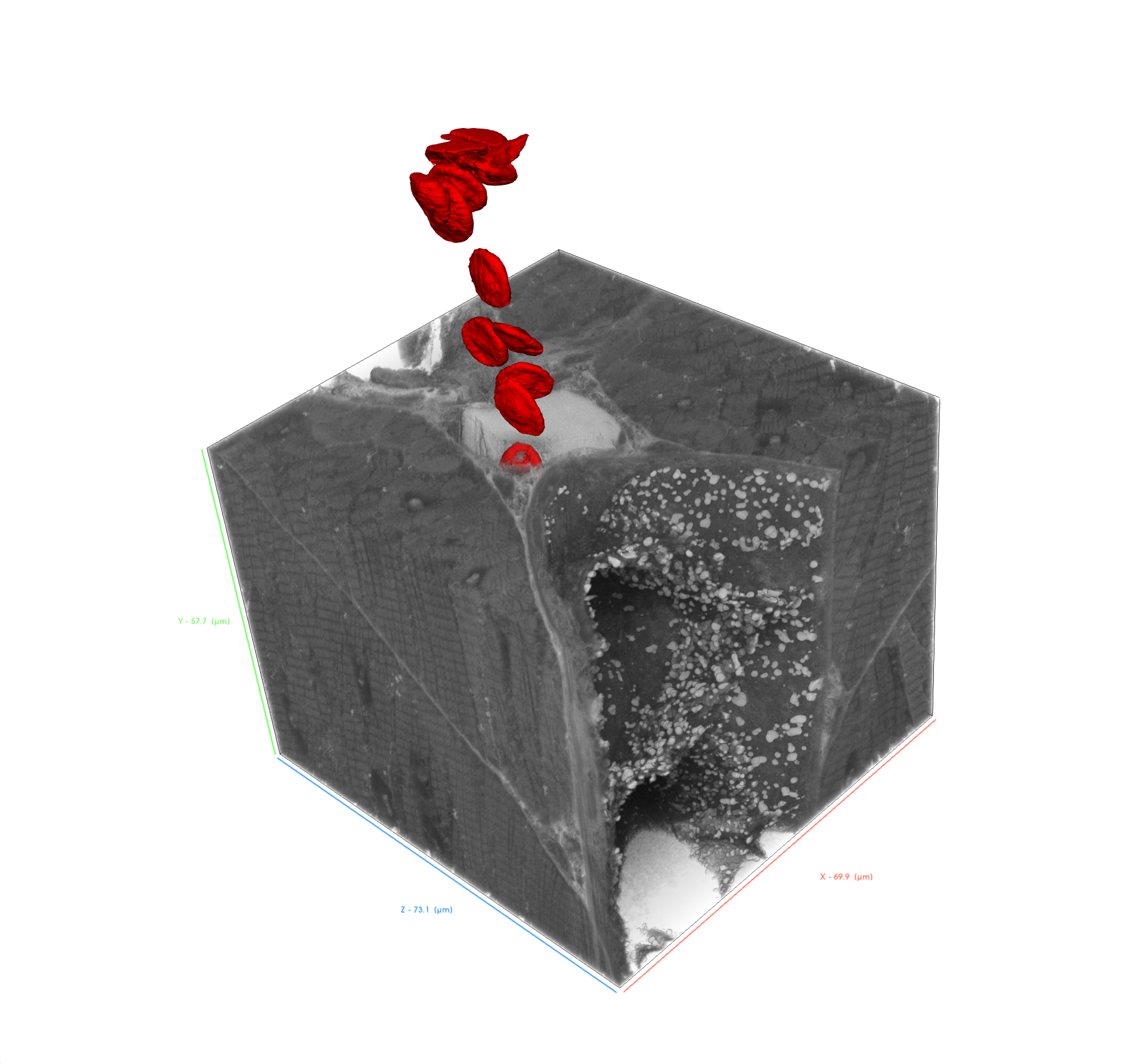
  • Ultra-high resolution SEM detection: visualize delicate biological structures with nanometer precision thanks to immersion optics

  • Fast, large-volume milling: process bigger sections more efficiently than with gallium FIB-SEM

  • Accurate and repeatable milling: ensure surface consistency and artifact-free imaging

SOLARIS-X2

GET IN Touch

Contact us

map

Where can you find us:

Tescan Brno
Libušina třída 21
623 00 Brno
Czech Republic

info@Tescan.com